


How Dental Plaque Biofilm Develops and Why It Drives Periodontal Disease
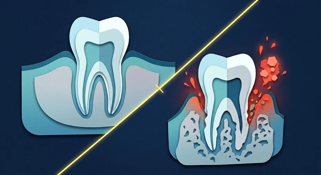
Bleeding on brushing, persistent bad breath and swollen gingival margins are among the most common reasons patients seek dental care.
Often the explanation is simple: plaque has stayed along the gingival margin and the tissue has become inflamed. Sometimes it is not. A patient has little visible plaque, yet the probe finds clinical attachment loss.
So how does a thin film on enamel contribute to the loss of the bone that supports the tooth? The answer goes beyond “poor brushing”. Plaque is a living, organised biofilm, and the host’s response to it largely shapes whether the outcome is gingivitis or periodontitis.
How Does Dental Plaque Form?
Plaque is often described as a soft deposit on the teeth. That is accurate, but it undersells it. It is a structured community of microorganisms living inside a protective matrix of their own making.
Formation begins within minutes of cleaning. Salivary proteins coat the enamel and form the acquired pellicle, which gives bacteria something to attach to.
Early colonisers, mainly Streptococcus and Actinomyces species, attach first. They then provide binding sites for other organisms through co-adhesion. As the biofilm thickens, oxygen falls in the deeper layers, which favours anaerobes.
Key point: Mature plaque is not simply more bacteria. It is a changing ecosystem in which organisms influence one another.
This topic sits at the core of UK BDS teaching, and it matters equally for hygiene, therapy and oral health science students, because biofilm control underpins prevention and periodontal assessment.
Why Does Plaque Affect Patients So Differently?
Two patients can carry similar plaque deposits. One has red, bleeding gingivae and nothing more. The other has periodontal pockets and radiographic bone loss.
Part of the explanation is the balance between the oral microbiome and the host response. Disease becomes more likely when environmental changes disturb that balance and favour a community associated with inflammation. This shift is called dysbiosis.
Two conditions need to be kept clearly apart:
- Gingivitis: inflammation of the gingiva without periodontitis-related clinical attachment loss. Plaque-induced gingivitis is generally reversible with effective plaque control.
- Periodontitis: destruction of the tooth-supporting structures, including periodontal ligament attachment and alveolar bone.
One point catches people out. A patient treated for periodontitis can still develop gingival inflammation around a reduced periodontium. That is not simple gingivitis. They remain a periodontitis patient and need ongoing monitoring.
Smoking complicates things too. Smokers can show less visible gingival bleeding despite higher periodontal risk, so a quiet-looking mouth is not necessarily a healthy one.
Whether you are revising alone or comparing your work with guidance from a Dentistry Assignment Writing Service UK, the same principle applies. Strong periodontal answers link susceptibility, microbial change and clinical measurements, rather than listing them side by side.
The Core Concepts
Microbial succession and the ecological plaque hypothesis
Organisms associated with periodontitis include Porphyromonas gingivalis, Tannerella forsythia and Treponema denticola. Naming them, though, does not explain the disease. The ecological plaque hypothesis proposes that changes in the local environment favour disease-associated organisms.
Here is a detail that adds real depth. As inflammation develops, gingival crevicular fluid (GCF) flow increases and its composition changes. The fluid brings protein-derived compounds into the pocket, including breakdown products that can act as nutrients.
These conditions may favour organisms suited to a protein-rich, inflammatory environment. That helps explain why biofilm and inflammation can become self-reinforcing, which goes beyond the usual “less oxygen, more anaerobes” explanation.
The host inflammatory response
The immune system recognises microbial components through pattern-recognition receptors, triggering inflammatory signalling and recruiting neutrophils and macrophages. Mediators such as interleukin-1 beta, TNF-alpha and prostaglandin E2 influence how intense and persistent the inflammation becomes. The problem arises when a protective response becomes excessive or poorly regulated.
How the tissues are destroyed
- Matrix metalloproteinases (MMPs) contribute to collagen breakdown in the periodontal connective tissue.
- RANKL promotes osteoclast formation and activity, driving alveolar bone resorption.
Key point: Bacteria do not simply eat away the bone. The damage reflects the interaction between microbial challenge and the host’s own inflammatory response.
Factors That Shape Disease
Biofilm control. Brushing may not reach every interproximal area, so interdental brushes or floss help. Crowding, overhanging restorations and poorly contoured margins make cleaning harder.
Smoking. It is associated with greater periodontal destruction and impaired healing, and it can reduce visible bleeding. Bleeding scores should never be used in isolation.
Diabetes. Poor glycaemic control is associated with increased periodontal susceptibility. Evidence suggests a bidirectional relationship, though the effect varies between individuals.
Periodontal measurements. Probing pocket depth runs from the gingival margin to the base of the pocket. Clinical attachment level (CAL) is measured from the cemento-enamel junction. A patient with gingival enlargement can have increased probing depths with no equivalent attachment loss, which is a classic route to misdiagnosis.
Turning the Science Into Clinical Reasoning
Follow the chain: biofilm formation → microbial shift → immune activation → tissue destruction → clinical picture.
Now compare two patients. Patient A has swollen papillae, visible plaque and bleeding on brushing, with no attachment loss. That is consistent with plaque-induced gingivitis. Patient B has deeper pockets, attachment loss and radiographic bone loss, which points towards periodontitis.
A practical tip for written work: when you mention any mediator, follow the pattern mediator → process → effect on tissue. For example: RANKL → osteoclast activation → alveolar bone loss.
For evidence, use peer-reviewed research and recognised textbooks. The 2017 World Workshop classification, published in 2018, is relevant because staging and grading shape how periodontitis is diagnosed and communicated today. Also separate laboratory evidence from clinical evidence, since a bacterium affecting a pathway in vitro does not show its contribution in every patient.
Mistakes That Cost Marks
- Treating all plaque bacteria as harmful. Disease reflects a shift in balance.
- Confusing gingivitis with periodontitis. Bleeding and swelling do not establish attachment loss.
- Blaming bacteria for all destruction. The host response contributes significantly.
- Treating plaque and calculus as identical. Calculus is mineralised plaque.
- Relying only on probing depth. Gingival enlargement can inflate it.
Bringing It Together
Plaque matures into an organised community, the environment shapes its composition, and the immune system responds. Sometimes inflammation stays within the gingiva. Sometimes it becomes prolonged and dysregulated, and connective tissue and alveolar bone are lost.
The real skill is not knowing that plaque causes inflammation. It is understanding why it develops, why it differs between patients and when it becomes destructive.
- Technology for Students
- Admissions & Applications
- Online Courses
- Career and Jobs
- Campus Life
- Drinks
- Film
- Fitness
- Food
- Παιχνίδια
- Gardening
- Health
- Κεντρική Σελίδα
- Literature
- Music
- Networking For Students
- Insights
- Party
- Religion
- Shopping
- Sports
- Theater
- Wellness
- Education News
- Business & Finance For Students
- Security, Law & Crime Education
- Insurance
- Science & Technology For Students
 Greek
 English
 Arabic
 French
 Spanish
 Portuguese
 Deutsch
 Turkish
 Dutch
 Italiano
 Russian
 Romaian
 Portuguese (Brazil)